

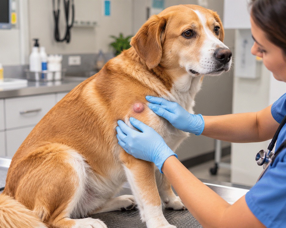
How fast tumors grow on dogs depends on the tumor type. A fatty lipoma may change little for long stretches, while a mast cell tumor can change size from day to day. Because a lump’s appearance can’t tell them apart, a needle sample is the usual first step to learn what a lump is.
Size alone doesn’t answer the question. Merck notes that mast cell tumors can mimic lipomas and that visual signs alone can’t establish a diagnosis, which is why testing comes before any decision about surgery.
How Fast Do Tumors Grow on Dogs? It Depends on the Type
According to the Merck Veterinary Manual’s guide to skin tumors in dogs, lipomas are benign fatty tumors, but some grow and can look identical to infiltrative lipomas or liposarcomas. Histiocytomas, common in dogs younger than about 3½ years, usually resolve on their own within 2 to 3 months. Mast cell tumors vary greatly in size and growth rate, and small ones may stay quiet for long periods before spreading. Our on-site diagnostics are part of how we work up a new lump.
Mast cell tumors can also swell and shrink. The American College of Veterinary Surgeons explains that their size can vary from day to day as granules inside the cells release histamine and other substances that inflame the surrounding tissue.
Is a Lump on My Dog Cancerous? How Vets Find Out
Merck states that telling a benign tumor from a cancerous one requires specialized knowledge and laboratory testing, and that a fine needle aspirate is needed to rule out tumors that mimic lipomas, including mast cell tumors. The ACVS notes that a needle aspirate diagnoses a mast cell tumor in almost all cases. If you’re asking whether a lump on your dog is cancerous, sampling is the step that answers it.
| Lump Type | Typical Behavior | What Testing Adds |
| Lipoma | Benign fatty tumor that can slowly enlarge | Rules out a mast cell tumor or liposarcoma that looks the same |
| Histiocytoma | Common in young dogs, often resolves in 2 to 3 months | Confirms the diagnosis before you wait it out |
| Mast cell tumor | Variable growth, size can change day to day | Grade and margins come from the removed tissue |
| Soft tissue sarcoma | Often invades nearby tissue but is less likely to spread | Guides how much surrounding tissue to remove |
For more on what causes these growths, see our guide to what causes a skin growth in dogs.
Mast Cell Tumor in Dogs: What the Numbers Show
A mast cell tumor in dogs is the most common type of skin tumor in dogs. The ACVS reports that these tumors make up 14 to 21 percent of all skin tumors diagnosed in dogs, and that Boxers and Boston Terriers account for about half of all cases. Other breeds listed include Pugs, Labrador Retrievers, Golden Retrievers, Beagles, and Schnauzers.
Merck notes that mast cell tumors larger than 3 cm at surgery are associated with shorter survival, and that tumors near mucous membranes, feet, or the lower body are more likely to spread. Because size at the time of surgery is linked to outcome, a confirmed mast cell tumor is worth discussing with your vet promptly.
What Lump Removal Surgery for Dogs Involves
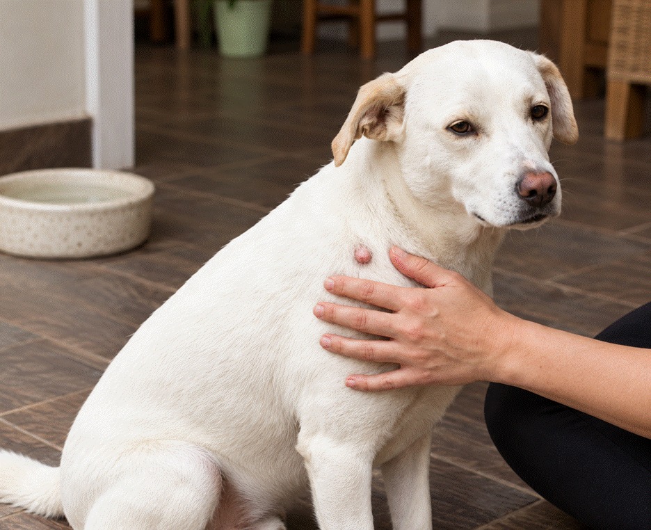
Lump removal surgery for dogs is planned around the diagnosis. The ACVS describes wide surgical margins of healthy tissue as necessary for mast cell tumors, and Merck notes that for suspected malignancies, tissue around the tumor is removed to lower the chance of leaving cells behind. Our tumor and mass removal guide explains the process from diagnosis to recovery.
Before surgery for a mast cell tumor, your vet may recommend staging tests. The ACVS lists aspirates of nearby lymph nodes, abdominal ultrasound, and chest X-rays as tests used to look for spread. After surgery, our guide to effective post-surgical care for dogs covers incision care at home.
What the Pathology Report Tells You
After removal, the tissue goes to a pathologist. Merck explains that the report describes the surgical margins this way:
● Dirty margins: tumor cells reach the edge of the removed tissue.
● Narrow margins: tumor cells are close to the edge but not at it.
● Wide or clean margins: tumor cells are far from the edge, so cells are unlikely to remain at the surgical site.
For mast cell tumors, the ACVS states that the grade can only be determined from the removed tissue and that it directly relates to prognosis.
Some reports include a genetic test. The ACVS notes that a c-KIT mutation is present in about 15 to 40 percent of mast cell tumors and is more common in higher-grade tumors, which carry higher recurrence rates and shorter survival times.
What Early Removal Can and Can’t Do
Early removal isn’t a guarantee. Merck reports that more than 70 percent of fibrosarcomas, a type of soft tissue sarcoma, return within a year of the first surgery, and that some dogs with mast cell tumors grow new tumors over time. Radiation and chemotherapy may still be needed after surgery, which the ACVS describes as decided case by case.
Spay timing is a common question for mammary tumors, and the evidence is limited. A 2012 systematic review in the Journal of Small Animal Practice judged the evidence that spaying reduces mammary tumor risk, and that the age at spaying matters, to be weak and not a sound basis for firm recommendations. If your dog is older, our senior pet health evaluations are one way to review overall health before surgery.
Staying Ahead of New Lumps
Feel over your dog when you groom, and note the date, location, and approximate size of anything new. A simple body map turns “I think it got bigger” into something your vet can compare.
At Kainer Veterinary Hospital, lump checks focus on getting a diagnosis first, followed by a removal plan if testing points to one. Contact us today to have a new or changing lump checked.
Sources
American College of Veterinary Surgeons, Mast Cell Tumors (2024). Supports mast cell tumor prevalence, breed risk, diagnosis by aspirate, margins, staging, grading, and c-KIT data.
Merck Veterinary Manual, Tumors of the Skin in Dogs (2018, updated 2026). Supports lipoma, histiocytoma, and sarcoma behavior, the fibrosarcoma recurrence figure, the size and location findings for mast cell tumors, and margin definitions.
Beauvais, Cardwell, and Brodbelt, Journal of Small Animal Practice (2012). Systematic review supporting the statement that evidence on spaying and mammary tumor risk is weak.
FAQs
Q: How fast do tumors grow on dogs?
A: It depends on the type. Lipomas may change little for long stretches but can enlarge, histiocytomas often resolve within 2 to 3 months, and mast cell tumors can change size from day to day.
Q: Are fatty tumors in dogs dangerous?
A: Lipomas are benign, but Merck notes that some grow and can look identical to infiltrative lipomas or liposarcomas. A needle sample is needed to rule out a mast cell tumor that mimics a lipoma.
Q: Is a lump on my dog cancerous?
A: Look and feel can’t answer that. A fine needle aspirate examined under a microscope is the usual first test, and a biopsy or removal may be needed for a full answer.
Q: Which dogs are more likely to get a mast cell tumor?
A: The ACVS reports that Boxers and Boston Terriers make up about half of all mast cell tumor cases, with Pugs, Labrador and Golden Retrievers, Beagles, and Schnauzers also listed.
Q: Does removing a tumor early improve the outcome?
A: For mast cell tumors, Merck notes that tumors larger than 3 cm at surgery are linked to shorter survival. Early removal can’t guarantee a cure, so follow-up still matters.