

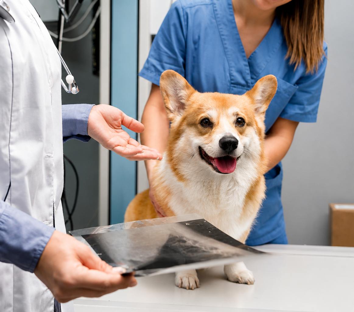
A pet scan for cardiology is usually an echocardiogram, an ultrasound of the heart that measures chamber size, wall thickness, valve function, and blood flow. For dogs with mitral valve disease, those measurements can decide whether medication starts, and in one large manufacturer-funded trial, starting at the right stage delayed heart failure by about 15 months.
The term can be confusing. A “PET scan” in human hospitals is a different technology. In veterinary heart care, the scan your vet means after hearing a murmur is almost always an echocardiogram, and this guide explains what it shows and what it can’t.
What a Pet Scan for Cardiology Shows
An echocardiogram builds a moving picture of the heart as it beats, showing how each chamber is sized, how the walls look, how the valves open and close, and how blood moves. A chest X-ray adds a view of the heart’s outline and the lungs. Our cardiology services include echocardiograms, electrocardiograms, and digital imaging, so the next step after a murmur can usually happen at the same hospital.
Which Pet Test for Heart Problems Fits Which Situation
Each pet test for heart disease answers a different question, so vets often combine them.
| Situation | Usual Test | What It Answers |
| New murmur in an older small-breed dog | Echocardiogram, often with chest X-rays | Whether the heart has enlarged enough to start medication |
| Coughing or fast breathing at rest | Chest X-rays | Whether fluid is building in the lungs |
| Irregular heartbeat or fainting | Electrocardiogram | What the heart’s electrical rhythm is doing |
| Older cat before anesthesia or IV fluids | Exam, NT-proBNP blood test, or echocardiogram | Whether hidden heart muscle disease is present |
Chest X-rays and the echocardiogram often work as a pair. For more on what X-rays reveal, see how a dog chest X-ray helps catch problems.
How a Scan Result Changes Treatment for Dogs With Valve Disease
Myxomatous mitral valve disease is the most common heart disease in dogs. The ACVIM consensus guidelines stage it from A, a dog at risk such as a Cavalier King Charles Spaniel, through D, advanced heart failure. Stage B dogs have a leaking valve but no signs of heart failure, and stage B splits in two.
In stage B1, the heart hasn’t enlarged. In stage B2, X-rays and an echocardiogram show enlargement that meets clinical trial criteria, and the guidelines recommend treatment to delay heart failure. No medication is recommended for stage B1, which is why measuring the heart matters.
The evidence for treating stage B2 comes from the EPIC trial, which was funded by Boehringer Ingelheim, the manufacturer of pimobendan. In 360 dogs, the median time to heart failure or cardiac death was 1,228 days with pimobendan versus 766 days with placebo, and median survival was 1,059 days versus 902 days. Our guide on why an echocardiogram matters for a dog with a heart murmur covers the murmur workup step by step.
Pets Worth Screening Earlie
Breed, species, and upcoming procedures change when a scan makes sense. Discuss screening with your vet for:
● Older small-breed dogs with a new murmur: the ACVIM guidelines identify breeds like the Cavalier King Charles Spaniel as at risk for mitral valve disease.
● Cats with or without a murmur: the ACVIM feline cardiomyopathy guidelines note that cats with heart muscle disease may have no murmur, and a murmur can occur in a cat with a normal heart.
● Maine Coon and Ragdoll cats: both breeds carry known genetic mutations linked to hypertrophic cardiomyopathy.
● Older cats before anesthesia, IV fluids, or long-acting steroid injections: the feline guidelines advise considering further heart testing first.
For the signs to watch for at home, see our article on hidden symptoms of heart disease in cats.
Watching Pet Heart Health at Home
A sleeping breathing count is a simple home tool for pet heart health. Research on sleeping respiratory rates reports that healthy dogs average fewer than 25 breaths per minute while asleep at home, and that dogs and cats with stable, well-controlled heart failure average under 30. Count breaths for 30 seconds while your pet sleeps in a cool room and multiply by two.
Skip counts taken after exercise or in the Conroe summer heat, since panting raises the number for reasons unrelated to the heart. A rate that stays above 30, or a steady upward trend, is a reason to call your vet. This is part of why early detection of heart disease depends on what owners track between visits.
What a Scan Can’t Tell You
A pet scan for heart problems stages the disease today. It doesn’t predict exactly when a pet will develop heart failure. The EPIC figures are medians, so half of the treated dogs reached the endpoint before 1,228 days and half after, and the trial only enrolled dogs that met its specific stage B2 criteria.
Treatment results in cats also depend on the stage found. For cats at higher risk of blood clots, the feline guidelines discuss medication to reduce clotting risk. A normal scan isn’t a lifetime clearance either, so recheck timing is set by your vet based on what the first scan showed.
Asking About a Scan
If your vet has mentioned a murmur, the useful question is what that murmur means. A scan answers it and gives your vet a baseline for every future visit.
Kainer Veterinary Hospital offers echocardiograms, electrocardiograms, and cardiac X-rays for dogs and cats. If your pet has a new murmur or breathing changes, contact us today to schedule a cardiac evaluation.
Sources
Keene et al., ACVIM Consensus Guidelines for Myxomatous Mitral Valve Disease in Dogs, Journal of Veterinary Internal Medicine (2019). Supports staging, at-risk breeds, and treatment recommendations by stage.
Boswood et al., The EPIC Study, Journal of Veterinary Internal Medicine (2016). Funded by Boehringer Ingelheim; supports the 1,228 versus 766 day and 1,059 versus 902 day results.
Luis Fuentes et al., ACVIM Consensus Statement on Feline Cardiomyopathies, Journal of Veterinary Internal Medicine (2020). Supports murmur limits in cats, breed risk, testing before procedures, and clot-risk medication.
Porciello et al., The Veterinary Journal (2016). Supports the sleeping breathing rate thresholds for dogs and cats.
FAQs
Q: What is the difference between an echocardiogram and a regular heart exam?
A: A regular heart exam uses a stethoscope to find murmurs and irregular beats. An echocardiogram is an ultrasound that shows chamber size, wall thickness, valve function, and blood flow, so it shows what’s causing the sound.
Q: Does my pet need sedation for a cardiac scan?
A: The scan is painless and requires lying still. Whether a calming medication is used depends on your pet’s temperament and health, and your vet will discuss it with you before the scan.
Q: Can a pet scan for cardiology tell me if my dog needs medication?
A: For mitral valve disease, yes. A pet scan for cardiology helps show whether a dog is in stage B1, where no medication is recommended, or stage B2, where the guidelines recommend treatment to delay heart failure.
Q: Which pets should be screened for heart disease even without symptoms?
A: Screening is worth discussing for older small-breed dogs with a new murmur, Maine Coon and Ragdoll cats, and older cats before anesthesia, IV fluids, or long-acting steroid injections.
Q: Can a pet scan for heart problems detect issues before symptoms appear?
A: Yes. A pet scan for heart problems can find heart enlargement and thickened heart muscle before coughing or breathing changes start, which is when treatment for stage B2 valve disease begins.