

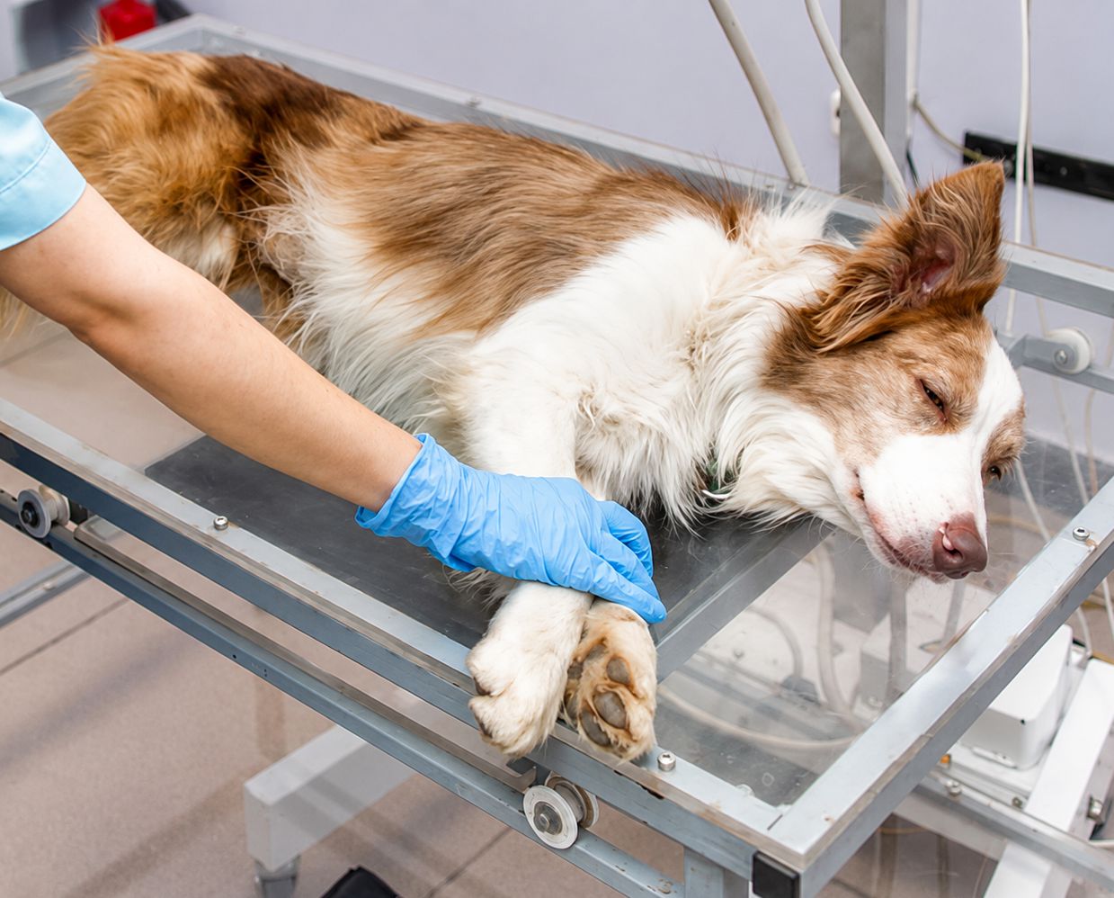
A dog chest x-ray produces a detailed image of the heart, lungs, major blood vessels, and the bones of the chest, without any incision. Veterinarians recommend it most often for a persistent cough, labored breathing, a new heart murmur, recent trauma, or to check whether cancer elsewhere has spread to the lungs.
Understanding the Dog Chest X-Ray
What a Chest X-Ray Actually Captures
A dog chest x-ray shows everything inside the chest cavity at once. Because the lungs, heart, blood vessels, and bone all sit at different densities, the resulting image shows a remarkable amount of detail about the size, shape, and condition of each structure in a single pass.
Why This Imaging Is So Widely Used
Chest imaging answers a wide range of clinical questions with one set of images: a veterinarian evaluating a cough is checking the lungs, one evaluating a heart murmur is checking the heart’s size and shape, and one managing a cancer diagnosis elsewhere is checking whether it has spread to the lungs. It is noninvasive, takes only a few minutes, and rarely requires an overnight stay, which is why it fits easily into a same-day visit alongside a physical exam.
What Owners Typically Notice Before the Recommendation
A cough lasting more than a few days, breathing that looks faster or more labored than normal, reduced energy, or a reluctance to lie down comfortably are common signs that prompt a veterinarian to look more closely at the chest.
How a Chest X-Ray Differs From a CT Scan or Ultrasound
A dog chest x-ray is usually the first imaging step because it is fast and widely available. A CT scan provides finer detail and can be necessary before a complex surgery or when a mass needs to be mapped precisely, while an ultrasound is better suited to evaluating the heart’s internal motion in real time.
Common Reasons a Dog Chest X-Ray Is Recommended
Persistent Coughing or Labored Breathing
A cough that lingers for more than a few days, or breathing that looks labored or unusually fast at rest, are common reasons a veterinarian recommends chest imaging. These signs can point toward several different conditions, from infection to fluid around the lungs to a mass.
Heart Murmurs and Suspected Heart Disease
When a veterinarian hears a heart murmur or other abnormal heart sound, chest imaging is often the next step to evaluate the heart’s size and shape and to look for early fluid buildup in the lungs, a sign of heart failure. This imaging complements rather than replaces other cardiac diagnostics, and the practice’s cardiology service covers how these tools work together.
Cancer Staging
For a dog already diagnosed with cancer elsewhere in the body, chest imaging is frequently recommended to check whether the disease has spread to the lungs, a process called staging. The tumor removal surgery for pets guide touches on how staging fits into the broader treatment picture.
Trauma and Injury
After a fall, a car accident, or any significant impact, chest imaging checks for injuries that are not visible from the outside, including a collapsed lung, fractured ribs, or bleeding within the chest cavity. Some of these injuries worsen over the hours following an impact rather than showing their full extent right away.
Pre-Anesthetic Screening for Certain Dogs
Some dogs, particularly seniors or those with a known heart or lung condition, benefit from a baseline chest x-ray before anesthesia for an unrelated procedure. Not every dog needs this extra step — a young, healthy dog undergoing a routine procedure typically does not need it, while a senior dog or one with a heart murmur benefits from the additional information it provides.
What the Images Can Reveal
| Finding on the Image | What It May Indicate |
| Enlarged heart silhouette | Possible heart disease, often followed by further cardiac evaluation |
| Increased density in lung fields | Possible pneumonia, fluid buildup, or bleeding within the lungs |
| A distinct mass or nodule | Possible tumor, which may require further imaging or a biopsy to characterize |
| Air outside the lung tissue | Possible collapsed lung, often related to recent trauma |
| Rib or spinal abnormalities | Fractures, old healed injuries, or less commonly a bone tumor |
A Normal Result Is Genuinely Useful Information
A dog chest x-ray that comes back normal is not a wasted step. Ruling out the lungs, heart, and chest structures as the source of a symptom narrows the list of remaining possibilities considerably.
Following a Finding Over Time
Some findings are not urgent on their own but are worth tracking, such as a small nodule that needs monitoring for growth or a heart that is only mildly enlarged.
What to Bring If Imaging Was Already Started Elsewhere
If your dog had chest imaging done at an emergency clinic or another practice before this visit, bringing along a copy of those images lets your veterinarian compare them directly against new films if repeat imaging is recommended.
How the Imaging Is Performed
How Chest Imaging Differs From Abdominal Imaging
A dog chest x-ray focuses on the heart, lungs, and rib cage, while abdominal imaging looks at the stomach, intestines, liver, and other organs further back in the body.
How Age Changes What Veterinarians Look For
A puppy’s chest x-ray is read with growth in mind, since developing structures can look different from a mature chest without indicating a problem. A senior dog’s images are reviewed with age-related changes in mind too.
Positioning and Sedation
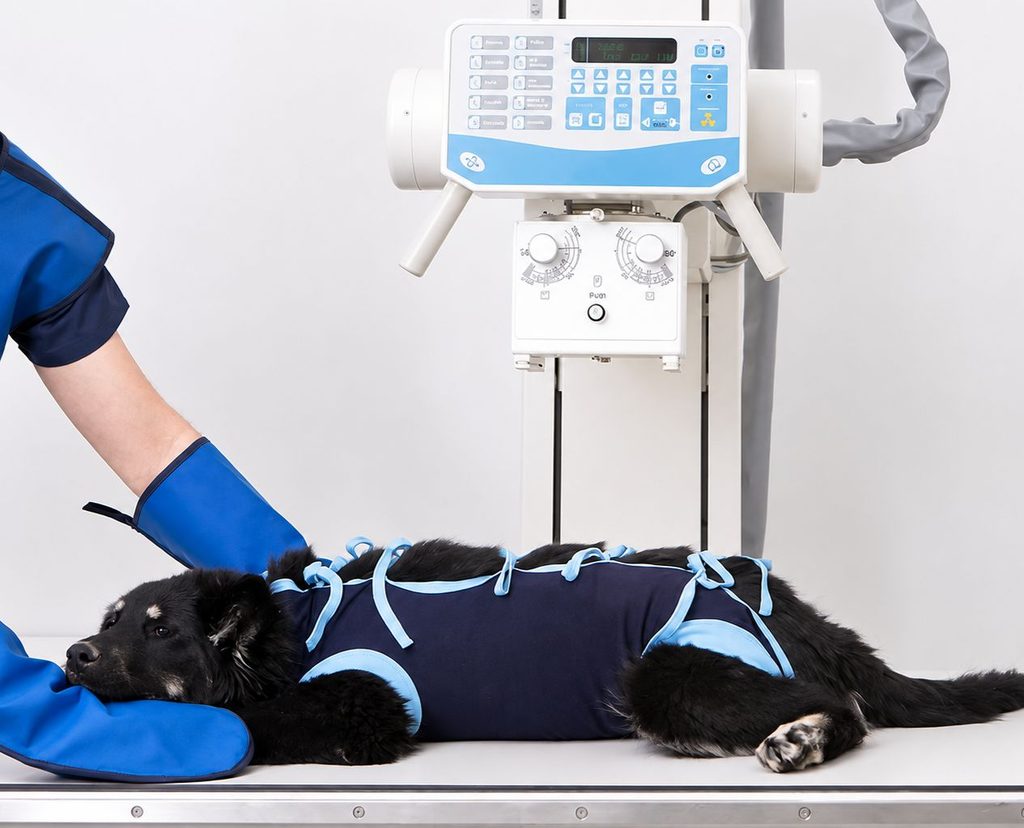
Most dogs are gently positioned for two or three different views of the chest. Some tolerate this while fully awake with gentle handling, while others, particularly those in pain or highly anxious, benefit from light sedation to get clear, motion-free images.
How Quickly Results Are Available
Most veterinary practices now use digital imaging, so the images are available for review within moments of being taken. On-site imaging capability is part of what the practice’s on-site diagnostics are built around.
What Happens After the Images Are Reviewed
Building a Complete Picture
A chest x-ray rarely stands alone as the only diagnostic step. The practice’s pet diagnostic tests guide is a helpful overview of how these tools fit together.
When a Specialist Referral Makes Sense
Some findings, such as a complex heart abnormality or a mass that needs a biopsy, benefit from the additional expertise of a board-certified specialist rather than being managed solely through general practice.
What Owners Can Do to Prepare
- Write down when the symptom started and whether anything seems to make it better or worse.
- Mention any recent injury or fall, even a minor one.
- Bring a list of current medications and supplements, which can matter if sedation is being considered.
- Ask directly whether sedation is recommended for your dog.
- Ask how and when you will receive the results.
What a Chest X-Ray Cannot Tell You
A dog chest x-ray is genuinely useful, but it is not the most sensitive tool available, and a page that only describes what it can find isn’t giving the full picture.
According to Dr. Eric Van Eerde, DVM, DACVR, a board-certified veterinary radiologist, chest x-rays often miss nodules smaller than about 7 to 9 millimeters, while CT scanning can detect nodules as small as 2 millimeters. That gap matters most for cancer staging, where a clean x-ray lowers the odds of spread to the lungs but does not rule it out completely.
Understanding What Comes Next for Your Dog
At Kainer Veterinary Hospital, Dr. Daniel Kainer and the diagnostic team provide on-site chest imaging to help identify what is affecting your dog’s heart, lungs, or chest, with same-day results in most cases. Contact the practice to schedule an evaluation if your dog is showing signs that concern you.
FAQs
Q: Why would my veterinarian recommend a dog chest x-ray?
A: A dog chest x-ray is commonly recommended for a persistent cough, labored breathing, a newly detected heart murmur, recent trauma, or to check whether a cancer diagnosed elsewhere has spread to the lungs.
Q: What can canine chest radiographs actually show?
A: Canine chest radiographs can reveal an enlarged heart, fluid or infection in the lungs, a mass or nodule, a collapsed lung, or fractures in the ribs and spine.
Q: Does my dog need to be sedated for a chest x-ray?
A: Many dogs tolerate a chest x-ray while awake with gentle positioning, though dogs who are anxious or in pain often benefit from light sedation to keep the images clear and motion-free.
Q: What does it mean if my dog’s chest x-ray comes back normal?
A: A normal result rules out the heart, lungs, and chest structures as the source of a symptom, which is genuinely useful even without an obvious finding.
Q: Where can I get diagnostic imaging for my dog in The Woodlands area?
A: Kainer Veterinary Hospital provides on-site chest imaging and diagnostic evaluation for pet families across The Woodlands, Conroe, Montgomery, and surrounding communities.